| Accession | GMS-171-1
URL copied to the clipboard |
| Title | Reconstructed nuclei from Xenopus laevis cell-free extract |
| Submit date | 2025-05-07 14:48:11 |
| Last update date | 2025-05-08 19:02:32 |
| Contact | Yuki HARA yukihara AT yamaguchi-u.ac.jp Yamaguchi University |
| Sharing | |
| Total file size | 311.55 KB |
| Keywords | Cell cycle Nuclear-scale Xenopus Pronucleus Cell-free reconstruction |
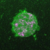
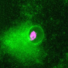
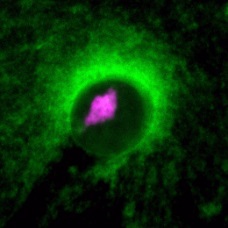
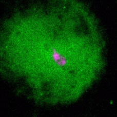
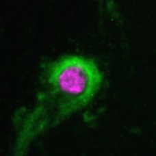
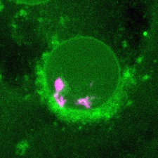
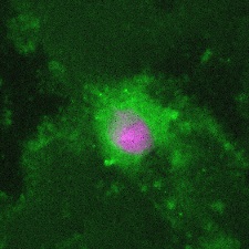
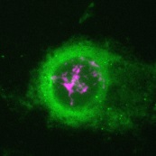
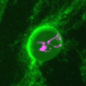
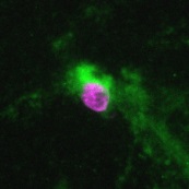
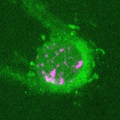
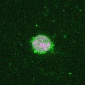
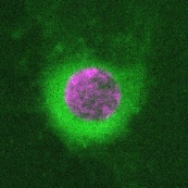
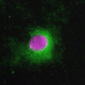
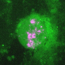
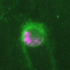
| Experiment type | The nuclei were reconstructed in the cytoplasmic extract from Xenopus laevis eggs with the demembranated sperm chromatin. We added RNase A at four different concentrations (100, 20, 5, and 1 μg/ml) into the extract just before incubation with sperm chromatin. After 40, 80, 120, and 180 min of incubation, we fixed the samples and prepared the imaging slides with Hoechst33342 (for visualizing DNA) and DiOC(6)3 (for visualizing the membrane). The images were taken by wide-field fluorescent microscopy equipped with a 40x objective. DNA and membrane were shown in magenta and green, respectively. The size of each image is 100 x 100 μm. The names of images are composed of the condition (control or RNase A concentration) and incubation time. |
| Summary | |
| Citation(s) |
| Organism | Xenopus |
| Cell (Tissue) | cell-free reconstruction |
| Protocol | |
| Data processing |


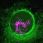

| [812] 100RNase_120min.jpg
the reconstructed nucleus at the presence of 100 µg/ml of RNase A, after 120 min of incubation image/jpeg 20.44 KB MD5: 4dd650a88eeed2c86c758ffe6ea3ea99 |
| [814] 100RNase_180min.jpg
the reconstructed nucleus at the presence of 100 µg/ml of RNase A, after 180 min of incubation image/jpeg 17.27 KB MD5: 478ee79434a295d016f91d90ea301bcc |
| [809] 100RNase_40min.jpg
the reconstructed nucleus at the presence of 100 µg/ml of RNase A, after 40 min of incubation image/jpeg 25.01 KB MD5: 4e21a35107f3ebeb38da1a3ee0a6d47f |
| [816] 100RNase_80min.jpg
the reconstructed nucleus at the presence of 100 µg/ml of RNase A, after 80 min of incubation image/jpeg 10.34 KB MD5: e64f346817ce0c93d7166546ac8ce683 |
| [805] 1RNase_180min.jpg
the reconstructed nucleus at the presence of 1 µg/ml of RNase A, after 180 min of incubation image/jpeg 26.85 KB MD5: 8e959726dba7c579f0a05b0b62597fb0 |
| [807] 1RNase_40min.jpg
the reconstructed nucleus at the presence of 1 µg/ml of RNase A, after 40 min of incubation image/jpeg 23.65 KB MD5: 7d13726bb9469494463cbbacda798767 |
| [810] 20RNase_120min.jpg
the reconstructed nucleus at the presence of 20 µg/ml of RNase A, after 120 min of incubation image/jpeg 9.04 KB MD5: 8be627bc543fd91be4d7f26ee86f5ee9 |
| [808] 20RNase_180min.jpg
the reconstructed nucleus at the presence of 20 µg/ml of RNase A, after 180 min of incubation image/jpeg 9.95 KB MD5: 74765968c67007219b197f4d9aafb4e2 |
| [813] 20RNase_40min.jpg
the reconstructed nucleus at the presence of 20 µg/ml of RNase A, after 40 min of incubation image/jpeg 8.23 KB MD5: e61eaa38aed621dd776d0e67cfeee366 |
| [811] 20RNase_80min.jpg
the reconstructed nucleus at the presence of 20 µg/ml of RNase A, after 80 min of incubation image/jpeg 10.08 KB MD5: c1aff6753244c6ee93d489992b51f012 |
| [806] 5RNase_120min.jpg
the reconstructed nucleus at the presence of 5 µg/ml of RNase A, after 120 min of incubation image/jpeg 15.80 KB MD5: 948750a01b42decb760277c873238254 |
| [817] 5RNase_180min.jpg
the reconstructed nucleus at the presence of 5 µg/ml of RNase A, after 180 min of incubation image/jpeg 11.22 KB MD5: 480eb54452f63abfa7f2eb0ffb1c62fe |
| [804] 5RNase_40min.jpg
the reconstructed nucleus at the presence of 5 µg/ml of RNase A, after 40 min of incubation image/jpeg 10.68 KB MD5: 34ff028fc02b773b8885b59aee142e60 |
| [800] 5RNase_80min.jpg
the reconstructed nucleus at the presence of 5 µg/ml of RNase A, after 80 min of incubation image/jpeg 14.81 KB MD5: 173e2619a507a324eb10f969df13a372 |
| [802] control_120min.jpg
the reconstructed nucleus (with dH2O), after 120 min of incubation image/jpeg 13.42 KB MD5: d29af1fd577b037033dd1149e816d521 |
| [801] control_180min.jpg
the reconstructed nucleus (with dH2O), after 180 min of incubation image/jpeg 12.95 KB MD5: f2d34fcd37e85f9867708bf71782cda6 |
| [818] control_40min.jpg
the reconstructed nucleus (with dH2O), after 40 min of incubation image/jpeg 7.99 KB MD5: e9287a53b94620249766921107fe70a3 |
| [815] control_80min.jpg
the reconstructed nucleus (with dH2O), after 80 min of incubation image/jpeg 8.40 KB MD5: 82599a4ec94aca066873c99b4c741ed8 |
| [799] 1RNase_120min.jpg
the reconstructed nucleus at the presence of 1 µg/ml of RNase A, after 120 min of incubation image/jpeg 29.75 KB MD5: 0ab50f2d01ea2c3fa94e6faeb2142de3 |
| [798] 1RNase_80min.jpg
the reconstructed nucleus at the presence of 1 µg/ml of RNase A, after 80 min of incubation image/jpeg 25.67 KB MD5: 1bc3368ddb162d1f2846b60b810ce6c0 |
