| Accession | GMS-65-6
URL copied to the clipboard |
| Title | Condensed but liquid-like domain organization of active chromatin regions in living human cells |
| Submit date | 2023-08-30 13:41:46 |
| Last update date | 2023-08-30 13:45:54 |
| Contact | GMSuite GENOMEMODALITY gmsuite AT hgc.jp The University of Tokyo |
| Sharing | |
| Total file size | 22.41 MB |
| Keywords | Live-cell imaging MSD Brownian dynamics |
| Experiment type | Single-nucleosome imaging Brownian dynamics simulation of chromatin next-generation sequencing |
| Summary | In eukaryotes, higher-order chromatin organization is spatiotemporally regulated as domains, for various cellular functions. However, their physical nature in living cells remains unclear (e.g., condensed domains or extended fiber loops; liquid-like or solid-like). Using novel approaches combining genomics, single-nucleosome imaging, and computational modeling, we investigated the physical organization and behavior of early DNA replicated regions in human cells, which correspond to Hi-C contact domains with active chromatin marks. Motion correlation analysis of two neighbor nucleosomes shows that nucleosomes form physically condensed domains with ~150-nm diameters, even in active chromatin regions. ...... [read more: https://pubmed.ncbi.nlm.nih.gov/37018405/ ] |
| Citation(s) | Nozaki T, Shinkai S, Ide S, Higashi K, Tamura S, Shimazoe MA, Nakagawa M, Suzuki Y, Okada Y, Sasai M, Onami S, Kurokawa K, Iida S, Maeshima K. Condensed but liquid-like domain organization of active chromatin regions in living human cells. Sci Adv. 2023 Apr 5;9(14):eadf1488. doi: 10.1126/sciadv.adf1488. Epub 2023 Apr 5. PMID: 37018405; PMCID: PMC10075990. https://pubmed.ncbi.nlm.nih.gov/37018405/ |
| Organism | Human |
| Cell (Tissue) | HeLa |
| Protocol | HeLa cells were cultured in Dulbecco’s modified Eagle’s medium (DMEM) (D5796-500ML; Sigma-Aldrich) supplemented with 10% fetal bovine serum (FBS) (FB-1061/500; Biosera) at 37°C in 5% CO2. For single-nucleosome imaging, cells were plated onto glass-bottomed dishes (3970-035; Iwaki) treated with poly-l-lysine (P1524-500MG; Sigma-Aldrich). Before microscopy imaging, the medium was replaced with DMEM lacking phenol red (21063-029; Thermo Fisher Scientific) and supplemented with 10% FBS. |
| Data processing |
|
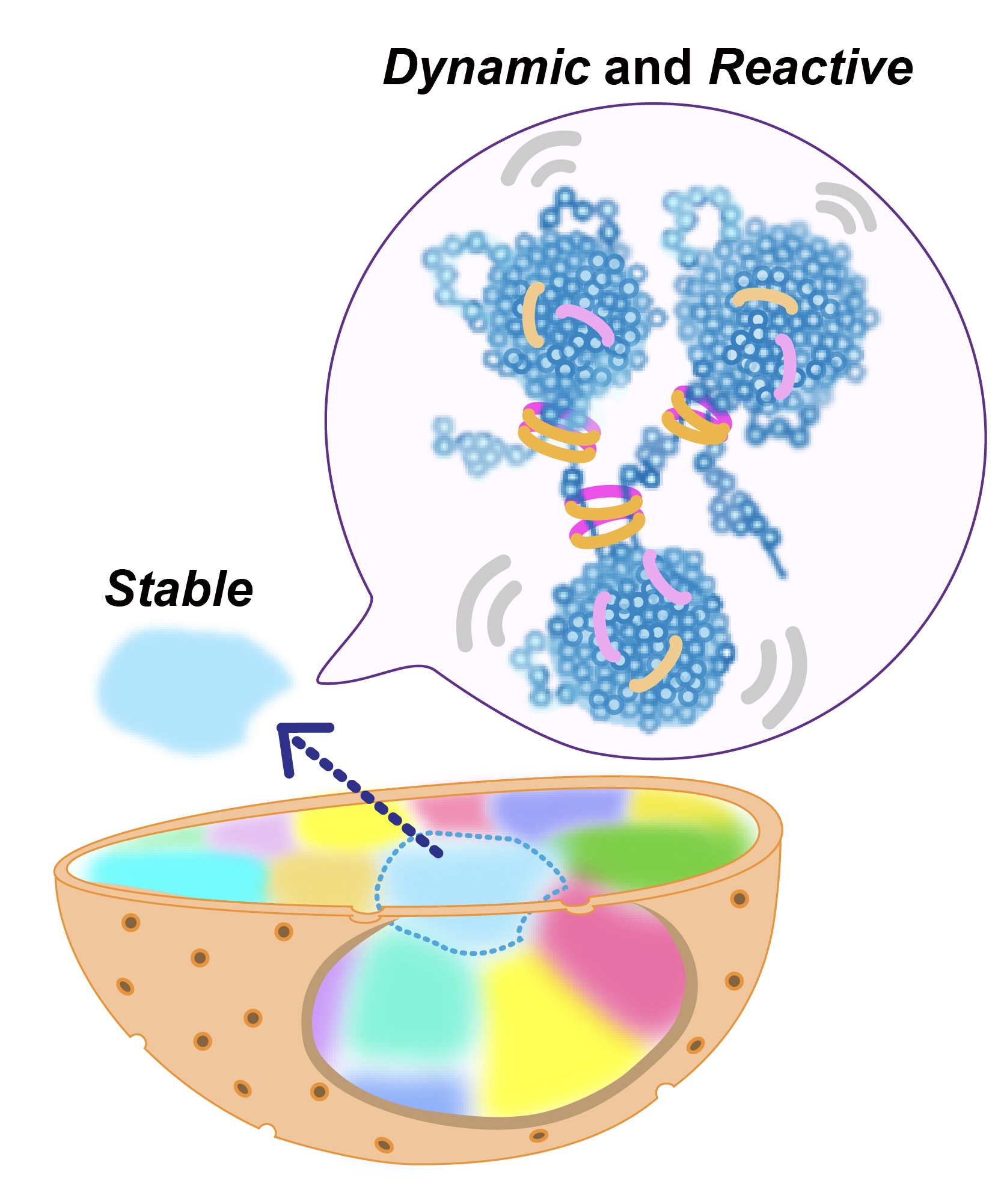
| [401] Figure8_220129.jpg
Nucleosomes (blue sphares) in active chromatin regions form condensed domains with local nucleosome contacts and cohesin (rings), where nucleosomes fluctuate. Each interphase chromosome (highlighted as different colors) is stably occupied in the nucleus image/jpeg 435.77 KB MD5: 053b2108e3f9c0adf2a8edf682075323 |
| [402] Final5_copy.mp4
Dual-color imaging of two neighbor nucleosomes reveals that nucleosomes in euchromatin form condensed domains with ~150 nm diameter. Nucleosomes locally fluctuate in the condensed domain like a liquid video/mp4 2.26 MB MD5: 97bcbdbc78c7c1009eeb2cfbb68abd0b |
| [405] Supplemental_MovieS10.mp4
A computational simulation of chromatin Brownian dynamics video/mp4 6.43 MB MD5: 04a6e955a666d6564553a38c1eb16dde |
| [407] Supplemental_MovieS11.mp4
A computational simulation of chromatin Brownian dynamics video/mp4 2.99 MB MD5: cf0f53168da452920b7efdf2ab8af1b8 |
| [408] Supplemental_MovieS12.mp4
A computational simulation of chromatin Brownian dynamics video/mp4 4.09 MB MD5: 001f70a13666768663a890d0b4128b42 |
| [406] Supplemental_MovieS13.mp4
A computational simulation of chromatin Brownian dynamics (cohesin KO) video/mp4 1.95 MB MD5: 342cca712d5069d359a325c72e7c14bf |
| [404] Supplemental_MovieS8.mp4
A computational simulation of chromatin Brownian dynamics video/mp4 3.05 MB MD5: 64bd0cd610f6e5df3ee50dbc84dd18ce |
| [403] Supplemental_MovieS9.mp4
A computational simulation of chromatin Brownian dynamics video/mp4 1.20 MB MD5: 16aa057ef3145220c63ccce9a010624f |
